Beyond the limit
31 May 2021
Smaller than light allows: Physicist Ralf Jungmann can also visualize individual proteins using conventional microscopes, bringing a new transparency to the nanoworld.
31 May 2021
Smaller than light allows: Physicist Ralf Jungmann can also visualize individual proteins using conventional microscopes, bringing a new transparency to the nanoworld.

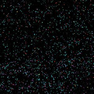
The DNA-PAINT method allows biomolecules to be identified under the light microscope. | © AG Jungmann
Physics isn’t kind to microscopy – at least not when it comes to optical insights into the depths of the nanocosmos. Since light, due to its wave nature, doesn’t always propagate in a straight line and can be deflected from its course by the tiniest of obstacles, the resolution of an optical microscope cannot be increased at will. There is an upper limit. A hard limit.
As German physicist Ernst Abbe calculated as far back as the late 19th century, that limit is approximately half the wavelength of visible light: 200 nanometers, or 200 millionths of a millimeter. Everything below that — whether viruses or proteins, which make up so many components of living cells and perform a wide range of control, communication and transportation tasks — remains hidden from the microscope. That’s what physics dictates. But physicists wouldn’t be physicists if they didn’t try to outfox physics. Like Ralf Jungmann. A professor of experimental physics at Ludwig-Maximilians-Universität in Munich (LMU), Jungmann has set himself the goal of focusing his microscope on individual molecules — despite any supposed limits. He wants to use super-resolution microscopy to visualize what goes on in the nanoworld of cells. He wants to observe how life functions at its most basic level. To do this — and to circumvent the laws of physics — he uses flashing probes, fragments of DNA and fluorescent light.
Super-resolution using fluorescence isn’t fundamentally new. In 2014, the Royal Swedish Academy of Sciences awarded the Nobel Prize in Chemistry to German physicist Stefan Hell and others for this method. A Director at the Max Planck Institute for Biophysical Chemistry in Göttingen, Germany, Hell developed a method in which interesting aspects of a cell are labeled with fluorescent molecules. One laser activates these self-luminous substances, while a second laser switches almost all of the light off again — with the exception of one tiny point, far smaller than Abbe’s limit. But this method is relatively complex. Not only does it require two lasers, but the sample also has to be approached and imaged point by point, as if being scanned.
Ralf Jungmann is pursuing a different approach. Easier and faster, or at least that’s what it promises: “Our technique is meant to be so simple that it can one day be used in any standard laboratory in the world,” says the biophysicist. At the core of this method are tiny DNA-based probes that attach to proteins or other cellular molecules and label them. When irradiated with light, these probes light up like the hands of a watch in the dark, thus revealing the position of the labeled object. But that’s not all: akin to a barcode that gets scanned at the supermarket checkout, the DNA probes also reveal which protein is being observed.
Jungmann named his technique DNA-PAINT. It sounds a bit like nanoart, but it has a very tangible background: The physicist’s labeling probes consist of a single strand of DNA. The exact complement of this DNA strand is floating in the cellular solution under the microscope. It is also bound with a fluorescent dye.
If this free-floating dye molecule comes into contact with the labeling probe, the two complementary DNA strands can unite to form a short double-helix structure. The dye remains at the desired location for a certain period, usually a few tenths of a second, before the bond dissociates again and the molecule disappears in the liquid.
Illuminating this with suitable light under the microscope yields a distinctive image: the dye molecules zipping around are hardly noticeable; they are moving too quickly to provide a clear fluorescence signal. However, as soon as it docks to the probe and comes to rest, it glows intensely and becomes noticeable. Then it sets off again and becomes invisible. This makes the labeling probe appear to flash like a lighthouse — and reveals the location of the protein to be analyzed.
This flashing offers the physicists several advantages: Labeling molecule that luminesce constantly would be of no benefit. These points of light would be subject to Abbe’s limit — like all objects under a microscope. However, if the point flashes, the individual images can be run through clever algorithms to localize the flash more precisely. Jungmann compares this trick with looking at several windows of a house at night. From a distance, if all windows are brightly lit, they look like a single light source. If, in contrast, the lights are alternately switched on and off, one can surmise the individual windows.
By using this ploy in his experiments, Ralf Jungmann, who also heads up the Molecular Imaging and Bionanotechnology research group at the Max Planck Institute of Biochemistry in Martinsried, was able to achieve a resolution of about five nanometers — 40 times better than Abbe’s limit. Five nanometers is equivalent to the size of a protein e.g. a cell-surface receptor — precisely what the researchers want to visualize.
The DNA probes offer yet another advantage: To visualize multiple protein types simultaneously, fluorescence microscopy currently uses different dye molecules for the individual proteins. However, the colors of the fluorescent light need to differ perceptibly, which limits the choice of suitable dyes. “As a result, only four or five different proteins can be observed at once,” says Jungmann. “But we want to look at hundreds or thousands of protein species.”
Dyes that vary in how frequently they dock to their targets are set to make this possible. If, for example, a labeling probe with five docking sites is attached to protein A, and a probe with ten docking sites to molecule B, B will get twice as many visits from the dyes floating around. Consequently, molecule B will also flash twice as fast. “In this way, every target molecule gets a characteristic flash code,” says Jungmann. This should make it possible to observe hundreds of proteins in 15 or 20 minutes.
Our technique is meant to be so simple that it can one day be used in any standard laboratory in the world.Prof. Dr. Ralf Jungmann, Professor of experimental physics at LMU. Head of the Molecular Imaging and Bionanotechnology research group at the Max Planck Institute of Biochemistry, Martinsried
The labeling probes are the biggest challenge here. Researchers typically use antibodies for this — parts of the immune defense that attack specific proteins. The problem with this is that antibodies are about three or four times the size of the target proteins they are supposed to label. If the antibodies start flashing once a dye docks to them, they flag only their own position and not that of the much smaller protein. The images become imprecise.
Jungmann and his team therefore set out to find better, smaller probes. They found them in aptamers — artificially produced DNA molecules that, like antibodies, can attach to certain proteins. From a pool of randomly generated aptamer sequences, they filter out the ones that bind particularly well with the desired proteins. Then the sequence of their DNA building blocks is determined, which forms the blueprint for future aptamer probes that can then be systematically produced in the lab.
This is particularly interesting for the DNA-PAINT technique. Since the docking sites for the dyes also consist of DNA, the blueprint for the aptamers merely needs to have a few DNA sequences added and voilà — a finished blueprint for a complete probe. This can then be produced by a DNA synthesizer, much like with a printer.
“Our goal was always to pack as much intelligence into the probes as possible, making it possible to forgo complex microscopes,” says Ralf Jungmann. Accordingly, the instruments the biophysicists use to test DNA-PAINT in the lab in Munich are commercially available fluorescence microscopes — supplemented by a laser that serves as the light source to make the labeling probes luminesce. According to Jungmann, this technique enables them to produce high-resolution images of the nanoworld with relatively modest equipment.
Probes are already commercially available, as well. Massive Photonics, a start-up co-founded by Ralf Jungmann and located in Gräfelfing, in Upper Bavaria, is selling the first kits for DNA-PAINT. Their aim, according to Jungmann, is to establish the technique in normal biology labs in the medium term — and that can be done only with clever, simple solutions: “If we were to tell laboratory operators that they first have to purchase a microscope for a million euros or hire two physicists two build such an apparatus, the entry barrier would definitely be too high,” says the LMU physicist.
Low barriers are also aimed at helping to make the desired applications for the new technology a reality. The researcher is particularly interested in cell-surface receptors — special proteins that facilitate communication between the cell interior and the outside world. Studying these would not only be interesting for basic research; the medical field and new drug development could also benefit from a better understanding of these kinds of processes. Many cancer drugs, for example, are designed to block cell-surface receptors, in the hope that doing so will suppress tumor signals.
But DNA-PAINT could also help to systematically examine tissue samples for traces of disease. “Our technology could be used to look at 10 or 20 cancer markers simultaneously, whereas it is currently possible to visualize just two or three markers,” says Jungmann.
All of that is still being researched. The physicists have, however, taken an important first step: they have shown that DNA-based microscopy can successfully overcome the supposed limits of their discipline.
Text: Alexander Stirn
Prof. Ralf Jungmann is a professor of experimental physics at LMU and heads up the Molecular Imaging and Bionanotechnology research group at the Max Planck Institute of Biochemistry in Martinsried. Born in 1981, Jungmann studied physics at Saarland University and the University of California, Santa Barbara in the US. He completed his PhD at the Technical University of Munich and was a postdoc at the Wyss Institute at Harvard University, Boston, USA. He led an Emmy Noether Independent Junior Research Group sponsored by the Deutsche Forschungsgemeinschaft, “DFG” (German Research Foundation), and was awarded a prestigious European Research Council Starting Grant.
Read more articles of the current issue and other selected stories in the online section of INSIGHTS/EINSICHTEN. Magazine.